Home » Without Label » Back Of Neck Anatomy : Triangles Of The Neck Anatomy Borders And Contents Kenhub : The occipital bone surrounds a large opening known as the foramen magnum.
Back Of Neck Anatomy : Triangles Of The Neck Anatomy Borders And Contents Kenhub : The occipital bone surrounds a large opening known as the foramen magnum.
Back Of Neck Anatomy : Triangles Of The Neck Anatomy Borders And Contents Kenhub : The occipital bone surrounds a large opening known as the foramen magnum.. Head and neck anatomy focuses on the structures of the head and neck of the human body, including the brain, bones, muscles, blood vessels, nerves in a newborn, the junction of the paritial bones with the frontal and occipital bones, form the anterior (front) and posterior (back) fontanelle, or soft spots. Browse 3,096 anatomy of neck and shoulder stock photos and images available, or start a new search to explore more stock photos and images. Related posts of anatomy of the back of the neck anatomy of respiratory system. Jugularis posterior) begins in the occipital region and returns the blood from the skin and superficial muscles in the upper and back part of the neck, lying between the splenius and trapezius. These muscles are mainly responsible for the movement of the head in all directions.
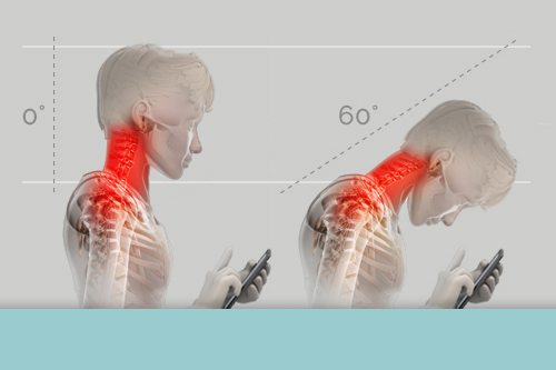
Think of it like a jigsaw puzzle, all the pieces fit in together and are required to get the full picture as to how it works. The occipital bone surrounds a large opening known as the foramen magnum. They ultimately drain into the deep lymph nodes. The back of the neck is mostly comprised of muscles, as well as the spine. Back pain is common and might be caused by a problem with a muscle.
Tips To Prevent Tech Neck And Other Pain From Technology Use from healthmatters.nyp.org These two ligaments connect and support the spine from the neck to the lower. Below the neck, holding the tooth into the bone, is the root of the tooth. Jugularis posterior) begins in the occipital region and returns the blood from the skin and superficial muscles in the upper and back part of the neck, lying between the splenius and trapezius. In this section, learn more about the anatomy of the muscles of the neck. The neck muscles, including the sternocleidomastoid and the trapezius, are responsible for the gross motor movement in the muscular system of the head and neck. The neck is essentially a passageway for air, food, liquids, blood, and more to travel between the head and the rest of the body, through structures such as blood vessels, nerves, and lymph nodes, as well as the larynx, trachea, and esophagus. These muscles are mainly responsible for the movement of the head in all directions. Back of neck anatomy :
It is composed of three parts:
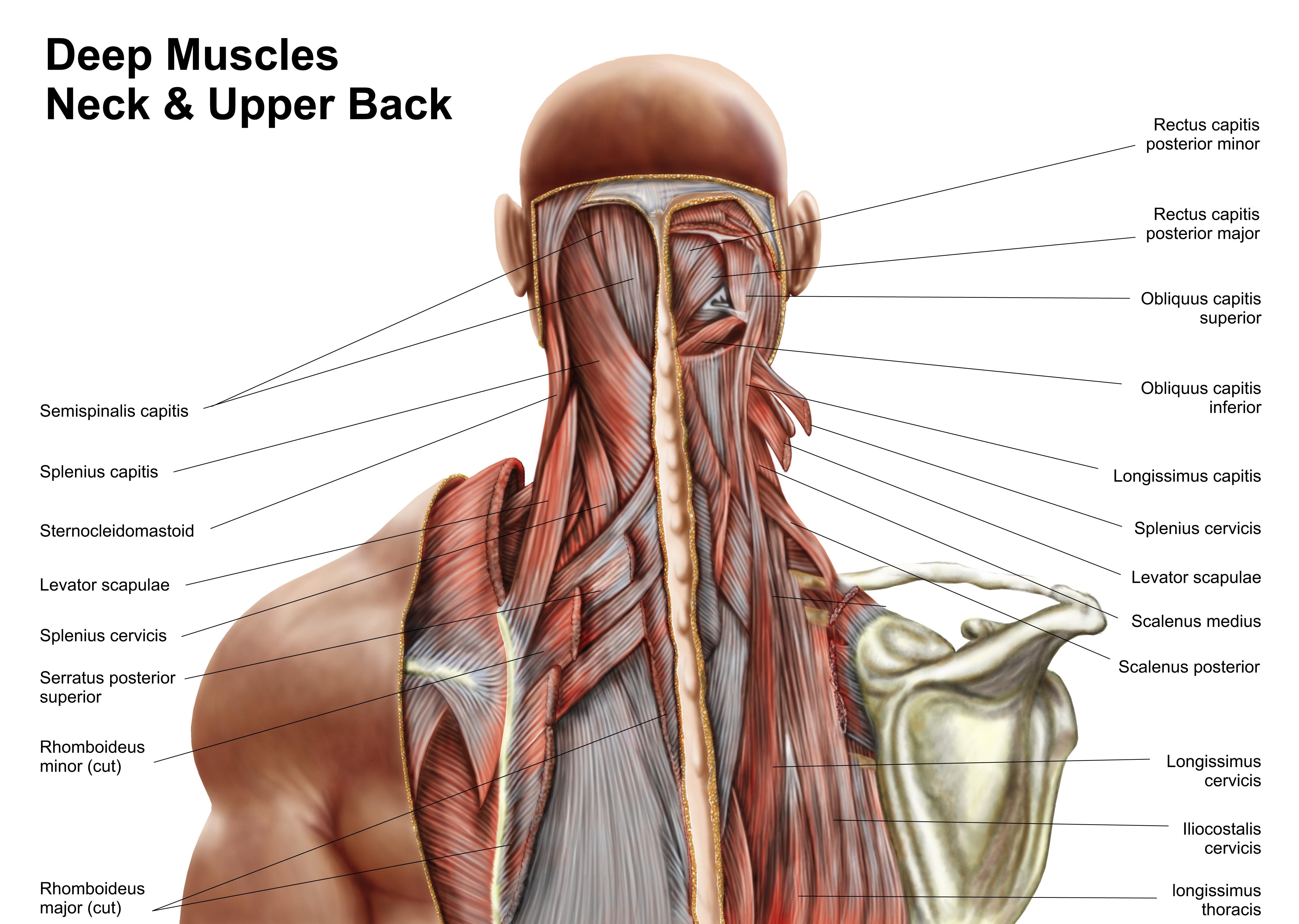
The trapezius muscle is a large muscle bundle that extends from the back of your head and neck to your shoulder. The top of the cervical spine connects to the skull, and the bottom connects to the upper back at about shoulder level. The majority of these nerves control the functions of the upper extremities and allow you to feel your arms, shoulder, and back of your head. They move the head in every direction, pulling the skull and jaw towards the shoulders, spine, and scapula. 12 photos of the anatomy of the back of the neck. Causes of neck pain and how to manage the pain in basic terms, the neck (cervical spine) joins the shoulders and chest to the head. In particular, the levator scapulae muscle is susceptible to injury. Neck anatomy explained the neck begins at the base of the skull and connects to the thoracic spine (the upper back). From this trunk, several vessels arise, which go on to supply the neck. See anatomy of the head and neck stock video clips. In this section, learn more about the anatomy of the muscles of the neck. Head and neck anatomy focuses on the structures of the head and neck of the human body, including the brain, bones, muscles, blood vessels, nerves in a newborn, the junction of the paritial bones with the frontal and occipital bones, form the anterior (front) and posterior (back) fontanelle, or soft spots. The muscles of the neck are muscles that cover the area of the neck .
Think of it like a jigsaw puzzle, all the pieces fit in together and are required to get the full picture as to how it works. The neck is essentially a passageway for air, food, liquids, blood, and more to travel between the head and the rest of the body, through structures such as blood vessels, nerves, and lymph nodes, as well as the larynx, trachea, and esophagus. The muscles of the neck are muscles that cover the area of the neck . The occipital bone is the only bone in your head that connects with your cervical spine (neck). The first branch of the thyrocervical trunk is the inferior thyroid artery.
Back Of Neck Anatomy Anatomy Drawing Diagram from image.pbs.org The occipital bone is a bone that covers the back of your head; Back of neck anatomy : Located at the back and side of the neck, the levator scapulae muscle connects the neck's cervical spine with the shoulder. They move the head in every direction, pulling the skull and jaw towards the shoulders, spine, and scapula. The anterior, and the posterior, triangles of the neck. The occipital bone surrounds a large opening known as the foramen magnum. This article gives an overview of the back's structure and its major muscles. It runs down the back part of the neck, and opens into the external jugular vein just below the middle of its.
Related posts of muscle anatomy back of neck piriformis muscle anatomy ultrasound.
These muscles give the sides of the neck their. It is composed of three parts: Choose from 500 different sets of flashcards about neck anatomy back neck upper on quizlet. An area called the occiput. The neck is connected to the upper back through a series of seven vertebral segments. Cervical spine anatomy video the cervical spine has 7 stacked bones called vertebrae, labeled c1 through c7. The neck is one of the most complex and intricate structures in our body and includes the spinal cord, which sends messages from the brain to the rest of the body. Related posts of anatomy of the back of the neck anatomy of respiratory system. It runs down the back part of the neck, and opens into the external jugular vein just below the middle of its. Neck anatomy explained the neck begins at the base of the skull and connects to the thoracic spine (the upper back). The anterior, and the posterior, triangles of the neck. The right and left subclavian arteries give rise to the thyrocervical trunk. The top of the cervical spine connects to the skull, and the bottom connects to the upper back at about shoulder level.
The posterior external jugular vein (v. Causes of neck pain and how to manage the pain in basic terms, the neck (cervical spine) joins the shoulders and chest to the head. These two ligaments connect and support the spine from the neck to the lower. The occipital bone is the only bone in your head that connects with your cervical spine (neck). Neck anatomy explained the neck begins at the base of the skull and connects to the thoracic spine (the upper back).
Levator Scapula Muscle And Its Role In Pain And Posture from www.verywellhealth.com The occipital bone is the only bone in your head that connects with your cervical spine (neck). They consist of 3 main groups of muscles: In addition, in this region we also find the major cranial and spinal nerves that connect the central nervous system to the organs, skin, and muscles of the head and neck. Head and neck anatomy focuses on the structures of the head and neck of the human body, including the brain, bones, muscles, blood vessels, nerves in a newborn, the junction of the paritial bones with the frontal and occipital bones, form the anterior (front) and posterior (back) fontanelle, or soft spots. Seek medical care if your neck pain is accompanied by numbness or loss of strength in your arms or hands or. The neck muscles, including the sternocleidomastoid and the trapezius, are responsible for the gross motor movement in the muscular system of the head and neck. Neck anatomy explained the neck begins at the base of the skull and connects to the thoracic spine (the upper back). Anatomy of respiratory system 12 photos of the anatomy of respiratory system anatomy and histology of the respiratory system ppt, anatomy and physiology of respiratory system with pneumonia, anatomy and physiology respiratory system worksheets, detailed anatomy of respiratory system, human anatomy respiratory.
The neck muscles, including the sternocleidomastoid and the trapezius, are responsible for the gross motor movement in the muscular system of the head and neck.
This muscle is controlled by the third and fourth cervical. In addition, in this region we also find the major cranial and spinal nerves that connect the central nervous system to the organs, skin, and muscles of the head and neck. The neck is connected to the upper back through a series of seven vertebral segments. The cervical spine, your neck, is a complex structure making up the first region of the spinal column starting immediately below the skull and ending at the first thoracic vertebra. In particular, the levator scapulae muscle is susceptible to injury. They start at the top of the neck and go down to the tailbone. These muscles give the sides of the neck their. See anatomy of the head and neck stock video clips. An area called the occiput. The neck muscles, including the sternocleidomastoid and the trapezius, are responsible for the gross motor movement in the muscular system of the head and neck. There are two main triangles; Located at the back and side of the neck, the levator scapulae muscle connects the neck's cervical spine with the shoulder. The superficial lymph nodes of the head and neck receive lymph from the scalp, face and neck.
:max_bytes(150000):strip_icc()/GettyImages-499158129-56a05f075f9b58eba4b0267f.jpg)
